Home
/ Chest Muscles Anatomy - 3d Rendered Medically Accurate Illustration Of The Chest Muscles Canstock / The pectoralis muscles are found on each side of your upper chest.
Chest Muscles Anatomy - 3d Rendered Medically Accurate Illustration Of The Chest Muscles Canstock / The pectoralis muscles are found on each side of your upper chest.
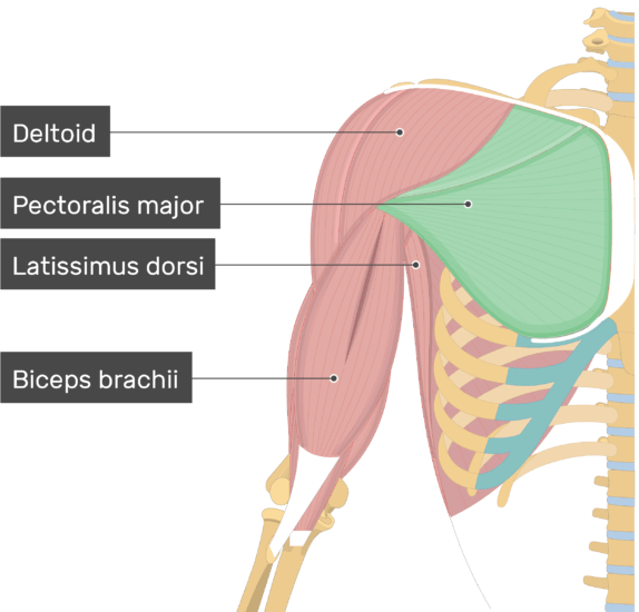
Chest Muscles Anatomy - 3d Rendered Medically Accurate Illustration Of The Chest Muscles Canstock / The pectoralis muscles are found on each side of your upper chest.. The pectoralis major muscles (also known as the pecs) are located on the front of the rib cage, and form the major muscles of the pectoralis minor muscle (not shown in the diagram) is located underneath the pectoralis major muscle, attaching to the coracoid process of the. The pecs attach to the humerus near the shoulder joint and originate on the breastbone in the center of the chest. You may also find triceps, lateral head brachialis, biceps brachii, latissimus dorsi, deltoid, acromion, clavicle, trapezius, 1st rib, clavicle, acromion, coracoid process, humerus, ulna, radius. There are around 650 skeletal muscles within the typical human body. These important muscles control many motions that involve moving the arms and head — such as throwing a ball, looking up at the sky, and raising your hand.
Choose from 500 different sets of flashcards about chest muscle anatomy on quizlet. The anatomical drawings were organized in a fairly classical manner to be easily used as a standard anatomical atlas. Muscles, connected to bones or internal organs and blood vessels, are in charge for movement. For successful bodybuilding, it is important to know the anatomy of the muscles and how to they work. Thigh magnetic resonance imaging the thigh has some of the body's largest muscles.
Pectoralis Major Muscle Attachment Action Innervation from www.getbodysmart.com The chest muscles are responsible for moving the arms across the body and up and down, as well as other movements like flexion, adduction, and rotation. Emg studies have shown that an *optimal. The anatomical drawings were organized in a fairly classical manner to be easily used as a standard anatomical atlas. Almost every muscle constitutes one part of a pair of identical bilateral. The anterior muscles of the trunk (torso) are associated with. As for the best bench angle to perform these with, this is something that will vary based on your anatomy. If you know where muscles attach and how. Anatomically, the chest is divided into two main regions:
As for the best bench angle to perform these with, this is something that will vary based on your anatomy.
It spreads out like a fan and covers the rib cage like an armor plate. You may also find triceps, lateral head brachialis, biceps brachii, latissimus dorsi, deltoid, acromion, clavicle, trapezius, 1st rib, clavicle, acromion, coracoid process, humerus, ulna, radius. It provides protection to vital organs (eg, heart and major vessels, lungs, liver) and provides stability for. The user can browse between different groups of images. Chest muscles is one of the large part muscles in your body that you also need to work out on aside from your arms, legs and core. Learn about chest muscles human anatomy with free interactive flashcards. The chest, or scientifically termed thorax, is located between the neck and abdomen, containing the thorax cavity and thorax wall. Using a rack for safety grab hold of the bar with a grip thats 15. The anatomical drawings were organized in a fairly classical manner to be easily used as a standard anatomical atlas. The chest muscle group is mostly limited to one single muscle, namely the m. Learn about chest muscle anatomy with free interactive flashcards. Anatomy, shoulder and upper limb, pectoral muscles. The pectoralis major, the pectoralis minor, and the serratus anterior.
Building chest muscles is a popular goal of training plan for those who are just starting their adventure with strength training. Learn anatomy faster and remember everything you learn. Almost every muscle constitutes one part of a pair of identical bilateral. These important muscles control many motions that involve moving the arms and head — such as throwing a ball, looking up at the sky, and raising your hand. You may also find triceps, lateral head brachialis, biceps brachii, latissimus dorsi, deltoid, acromion, clavicle, trapezius, 1st rib, clavicle, acromion, coracoid process, humerus, ulna, radius.
1 from Four main muscles in the pectoral region exert a force on the upper limb. Find out more about the individual muscles within the chest anatomy by clicking their respective links throughout this page. Learn anatomy faster and remember everything you learn. Choose from 500 different sets of flashcards about chest muscle anatomy on quizlet. The user can browse between different groups of images. Muscles, connected to bones or internal organs and blood vessels, are in charge for movement. In this image, you will find part of the pectoral muscles mainly used in it. To know why this is happening, it is worth knowing more about the anatomy of the chest muscles.
Thigh magnetic resonance imaging the thigh has some of the body's largest muscles.
This is a table of skeletal muscles of the human anatomy. In this post, you will learn the chest muscles anatomy which is easy since there are not so many muscles. Find out more about the individual muscles within the chest anatomy by clicking their respective links throughout this page. There are around 650 skeletal muscles within the typical human body. Almost every movement in the body is the outcome of muscle contraction. The anatomical drawings were organized in a fairly classical manner to be easily used as a standard anatomical atlas. In this article, we shall learn about the anatomy of the muscles of the anterior chest. Breathing, a vital body function, is also controlled by the muscles connected to the ribs of the chest and upper back. Anatomy, shoulder and upper limb, pectoral muscles. Four main muscles in the pectoral region exert a force on the upper limb. They are the pectoralis major, pectoralis minor, and the serratus anterior. Chest muscle anatomy the pectoralis major muscles also known as the pecs are located on the front of the rib cage and form the major muscles of the chest. It spreads out like a fan and covers the rib cage like an armor plate.
The pectoralis major has two anatomic sections or heads Anatomically, the chest is divided into two main regions: However, our primary focus is on the chest's anatomy or the chest's main muscles in this section. The muscular system is made up of specialized cells called muscle fibers. Building chest muscles is a popular goal of training plan for those who are just starting their adventure with strength training.
Anatomy Of Chest Muscles Bodybuilding Chest Muscles Images Stock Photos Vectors Shutterstock But I Believe That To Build Muscle You Have To Consciously Contract The Muscle That You Are Working from i0.wp.com Almost every movement in the body is the outcome of muscle contraction. Choose from 500 different sets of flashcards about chest muscle anatomy on quizlet. The chest, or scientifically termed thorax, is located between the neck and abdomen, containing the thorax cavity and thorax wall. The chest wall is comprised of skin, fat, muscles, and the thoracic skeleton. The pecs attach to the humerus near the shoulder joint and originate on the breastbone in the center of the chest. Deltoid muscles help you move your shoulders. The anatomical drawings were organized in a fairly classical manner to be easily used as a standard anatomical atlas. If you know where muscles attach and how.
Pectoral muscles are most predominantly associated with movement of the shoulders and arms.
The anatomical drawings were organized in a fairly classical manner to be easily used as a standard anatomical atlas. Chest muscle anatomy the pectoralis major muscles also known as the pecs are located on the front of the rib cage and form the major muscles of the chest. Chest muscles anatomy for bodybuilders. Breathing, a vital body function, is also controlled by the muscles connected to the ribs of the chest and upper back. Deltoid muscles help you move your shoulders. The anatomical drawings were organized in a fairly classical manner to be easily used as a standard anatomical atlas. Chest muscles is one of the large part muscles in your body that you also need to work out on aside from your arms, legs and core. The chest wall is comprised of skin, fat, muscles, and the thoracic skeleton. The pecs attach to the humerus near the shoulder joint and originate on the breastbone in the center of the chest. Primarily, there are three chest muscles involved in movement: Learn about each muscle, their locations & functional anatomy. This webpage presents the anatomical structures found on thigh mri. Choose from 500 different sets of flashcards about chest muscle anatomy on quizlet.
